The next step after covering off the basic skeleton and muscle systems of the body (the musculoskeletal system) is to focus on the bones that protect the system that regulates the muscles and other organs – the nervous system.
Sensitivity of Nerves
The nervous system is the most sensitive system of all the systems in the body due to the fundamental way the cells work – crudely speaking we could call it electricity as currents of charged particles are moving in and out as they communicate with each other. If we are going to be more precise it works on the sophistication of different charged ions being constantly pumped out and then allowed to flood into cells that allows specific messages to be communicated through the body (the effect of electricity on the body is to allow those ions to flood in causing muscles to contract, hormones to be released and other bad stuff if we want to stay well. It is very easy for the balance to be changed and that is why the nervous system is very sensitive to mechanical and chemical damage.
Mechanical Protection of the Nervous System – Armour
The mechanical protection of the nervous system is through two sets of bones: 1) the spinal column and 2) the skull. Together the spinal column and skull provide a hard armour preventing easy mechanical damage from going around the place.
The Spinal Column
The spinal column serves two main functions: 1) provides a strong column of bone to allow us to stand up on our feet with our heavy head balanced on top and 2) a centralised location for all the communicating nerves to connect to the main coordinating computer – the brain.
To provide flexibility to be able to run, stretch, turn and all other levels of agility the spine is composed of 32 different spinal bones or vertebra (plural vertebrae). These 32 bones are divided into five sections with a number of defined vertebra per section. In the first three sections the vertebra are separate to allow the body to twist and bend. As we move to the bottom of the spine the vertebrae fuse to provide a strong base that sits in the pelvis. The sections are as follows:
1) Cervical (neck) 7: C1-C7
2) Thoracic (chest) 12: T1-T12
3) Lumbar (abdominal) 5: L1-L5
4) Sacral (pelvis) 5: all fused)
5) Coccyx (tail) 4: all fused).

The three upper sections (cervical, thoracic, and lumbar) vertebrae not only provide movement they also provide a single pathway for nerves of the body to join up to the spinal column (through small bony openings called intervertebral foramina). As the nerves join to a single section of the spinal cord the body’s nervous system can be divided into connections into the spine. I’ll cover off how the nerves of the body connect to the central spinal cord when we look at the nervous system next.
This association between spinal vertebra and spinal cord is the foundation for spinal injury where an injury to the spinal column may result in an injury to the nerves associated to that area plus the nerves below it. For example an injury to lumbar vertebra L5 at the base of the spine can result in nerve signalling from the leg in forms of sensory (touch) detection and motor (muscle) movement. If the injury is further up, say at the top of the thoracic section (T1: neck and upper back) all the sections below this can be affected as this section is affects cuts-off signals going up and down the spine. Keeping the spinal vertebrae in good shape keeps the spinal cord in good shape and that is essential to bodily movement.
The Skull – Hard Hat of the Head
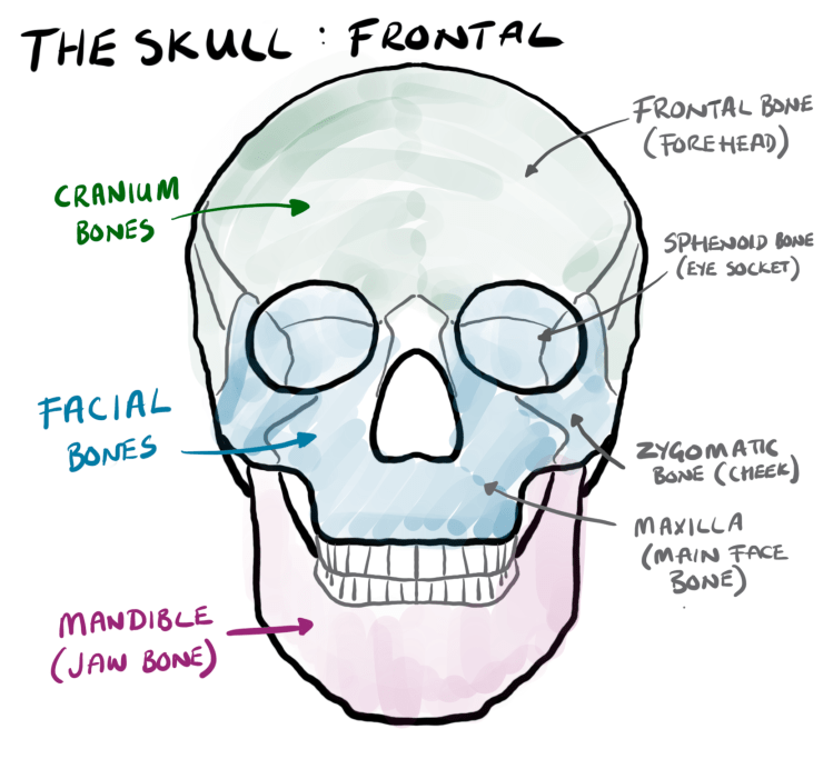
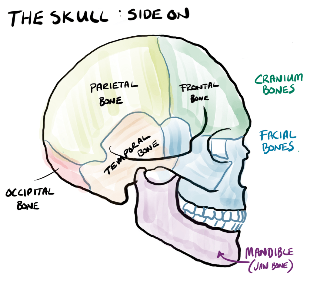
The skull is the hard bone that covers the brain and made up of two bones: 1) mandible or jaw bone and 2) the cranium. So if you need to order a skull at any time make sure you get both bits. I’m going to be a bit cheeky (pun intended) by dividing the cranium into facial bones (those that make up the face and eye sockets) and cranium bones (the ones that cover the brain). Antamotically there is no difference but it will help when we talk about the brain parts later on.
The Mandible – the Jaw Bone
The mandible is one bone that is attached to the side of the crainum via tendons the muslces that allow it to more up and down and side-to-side to chew or masticate (Greek for mastichan – chew/gnash with team) which gives the root of mandible (our reptile and bird friends can chew due to different jaw antonomy). This flexibility also allows for manipulation of the face to form different expressions.
Facial Bones
The cranium is more complicated but can be simplified to two many regions: 1) face (facial bones), brain (bones that cover the brain).
The facial bones are the ones that form the upper lip, nose all the way around to the side of the head. These bones create the openings for the eyes and the nose along with the top jaw or maxilla where the teeth join to the skull. Due to the complex nature of this part of the head there a lot of small bones (bones are really small plates that fuse or suture together) that make up the parts of the nose and eyes. Due to these smaller bones they are subject to breaking when the head gets hit. This can be an issue as the brain is right behind it along with a lot of blood vessels. Talking of blood vessels the nose structure is an interesting one. The nose is made of or the nasal bone and then cartilage that gives the nose it’s structure. When the nose gets ‘broken’ we are talking about the nasal bones at the top which hold the cartilage straight. If the nasal bones are not set straight that it were you get your wonky noses.
4 Main Cranium Bones
The rest of bones that make up the brain shell that provide that protective cover of the brain. A good place to start is the Atlas! One of the amazing things about the human head is it’s location and it’s weight. The brain is the central processing unit and the closer the brain is to the bodily sensors the better and that is why the eyes, ears, nose and tongue are right next to the brain for processing. By lifting the sensor as far off the floor as possible the further the body can see. Standing up-right and lots of sensors means a heavy head. To cope with that the skull head sits on top of the spinal column on the final bone called the Atlas Vertbra as in the Atlas from Greek mythology who has to hold up the heavens as he lost in the war of the Gods to Zeus (in reality it’s a mountain range in north Africa that would look like its the gap between the earth and the sky). The Atlas Vertebra provides the base for the cranium to sit on via hole called the foramen magnum (Latin: great hole!) formed within the occitipal bones. This hole provides the path for the nerves from the spinal cord into the brain not the blood vessels which come in from in front of the neck (carotid and vertebral arteries).

Once the cranium is sitting on top of the spinal column then four set bones cover the brain. From the front there is the frontal bone which makes up the forehead and eye sockets that fuse with the facial bones. Following the frontal bone on top of the cranium is the parietal bone (looking at it from the inside it provides the wall of back of the cranium). These come as a pair to cover the top of the head. For for the bones over the left and right hand side is the temporal bone that is above the ears. With the temporal and parietal bones making up the top and sides of the cranium the occitpital bone makes up the back and the base.
The four bones of the cranium: frontal, parietal, temporal, and occipital bones also make up the four lobes of the brain. Let’s move from the outside to the inside – the squishy stuff – the nervous system.
Step 3: Step3: Detect, Compute, Respond (or just Respond) – The Nervous System
